SMMART HTS — Automated Trace Evidence Microscopy System

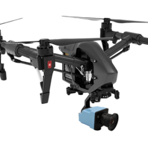
The SMMART HTS is the world’s first high-content screening microscope system purpose-built for forensic trace analysis. Developed by Spectricon and ranked first in a pan-European pre-commercial procurement (PCP) across leading forensic institutes, it has successfully passed site acceptance tests in major European national forensic labs.
What it does
Traditional trace evidence analysis is slow, subjective, and labor-intensive. The SMMART HTS automates this entire process — identifying, enumerating, and documenting millions of microscopic traces collected from crime scenes, in a fully unsupervised manner and at a speed 178× faster than manual examination. It handles the routine workload of a forensics lab so that experts can focus on high-value decisions.
before scan: after scan:

How it works
The system combines an advanced robotic microscope with a powerful machine learning platform. In a single automated scanning session, it captures a rich, multidimensional imaging dataset across a large sample stage (640 × 420 cm), using four complementary imaging modes:

Each scan produces a set of 40 multimodal images per field of view in just 5 seconds, generating up to 8 million spectra per spectral cube. The system can cover 3,600 fields of view in approximately 5 hours, entirely without operator intervention.
Intelligent analysis
The integrated software platform processes all acquired data — spectral, fluorescence, polarimetric, and morphological — to generate trace identification maps and enumeration tables automatically. Key capabilities include:
Why it matters
The SMMART HTS turns a process that was previously subjective and fatigue-prone into one that is objective, standardized, and scalable. It is suitable for both routine casework and research applications, is open to customization, and integrates seamlessly into existing forensic lab workflows as a turnkey solution.
| # | Specification | Value |
|---|---|---|
| 1 | Modes of operation |
|
| 2 | Sensor spatial resolution | 8 megapixels per image |
| 3 | Illumination modes |
|
| 4 | Acquisition data volume and speed | 40 images (multimodal image set); acquisition time 5 s per objective |
| 5 | Objectives | 2×, 5×, 15× (additional options possible) |
| 6 | Field of view (FOV) | 5 mm² (5× objective), magnification 265× |
| 7 | Autofocus | Electromechanical autofocus |
| 8 | Spectroscopy | 8 million spectra per spectral cube; spectra display and comparison on mouse hover |
| 9 | Polarimetry |
|
| 10 | Sample stage size | 640 × 420 cm (single unsupervised scan) |
| 11 | Sample scanning modes |
|
| 12 | Software |
|
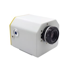
A cutting edge snapshot spectral imager acquiring up to 22,4K-level spatial resolution apectral images through a single,interchangeable lens
How would you define the ideal hyperspectral imager? you would agree that it shall acquire all spectral images instantly and at video rates and display all of them, together with the result of their analysis, i.e., spectral classification maps, in real-time!
Pushing the limits of what is possible in imaging science, Spectricon made this happen! MUSES9-SnS is the world’s first hyperspectral imager acquiring hypercube image packs at video rate and displaying both spectral images and classification maps side-by side and in real-time.
There is no tradeoff between spectral and spatial resolution, with the later reaching 4K levels!
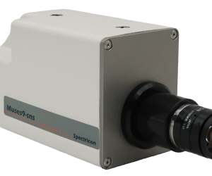
MUSES9-MS spectral camera addresses the need for combining spectral information from UV-VIS-NIR-SWIR, as a means for advancing the imaging and analytical capabilities of spectral imaging. Spectral imaging in this ultrawide spectral range has been proved to be very informative in a broad range of applications. Satellite cameras (AVIRIS, SENTINEL I & II) is a typical example of spectral imagers covering such a broad spectral range, with certain imaging bands being dedicated to specific identification/quantification targets.
Unlike satellite imaging, commercial spectral cameras are dedicated to operate within certain fractions of the 365-1700nm range, due to the restricted spectral sensitivity range of the available imaging sensor chips. This implies that more than one spectral imager is required if one wants to cover this wavelength range. However, this option is very costly, is of high volume and weight and the direct comparison of the spectral imaging data is far from being a straightforward process.
MUSES9-MS is an all in one spectral camera operating in the 365-1700nm spectral range. It effectively addresses the demand for an integrated, low cost, volume and weight, enabling the direct comparative analysis of the acquired, information-rich dataset.
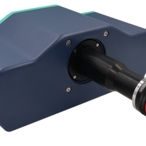
MUSES9-HS is a full frame, tunable filter-based hyperspectral imager, offering incomparable light throughput (94%) and tuning range (370-1000nm). Within a few dozen of seconds, MUSES9-HS automatically acquires a hypercube image stack comprising up to 126 (extendable to 315) sharply focused, 6.4-megapixel spectral images, collectively containing 6.4 million spectra.
The full-frame, tunable filter operation is clearly advantageous over line scanning or push broom traditional technologies, especially when examining static, two dimensional scenes. This is because bulky mechanical scanners are no longer required, to the benefit of portability, set up simplicity, versatility and direct adaptability to all kinds of lenses, microscopes and telescopes.
![]() +65 6748 5517
+65 6748 5517
![]() info@jm-vistec.com
info@jm-vistec.com
![]() 10 Kaki Bukit Ave 1, #07-06, Singapore 417942
10 Kaki Bukit Ave 1, #07-06, Singapore 417942